Keratoconus is a non-inflammatory corneal disease that causes the cornea to thin and gradually change shape. Normally, the cornea is spherical, but in keratoconus, it takes on a conical shape, leading to impairment of the eye’s optical system.
Keratoconus is a type of corneal dystrophy and often has a genetic predisposition. According to current data, inheritance is autosomal dominant with partial penetrance. The incidence of familial cases reaches approximately 10%.
The disease is associated with a number of systemic conditions, including Down, Marfan, and Turner syndromes, as well as atopic diseases, osteogenesis imperfecta, and mitral valve prolapse. Furthermore, it is often associated with other ophthalmological pathologies.
In practice, this means the disease requires comprehensive ophthalmological monitoring. Early diagnosis plays a key role in choosing treatment strategies and can slow progression in the early stages.
How does keratoconus manifest itself?
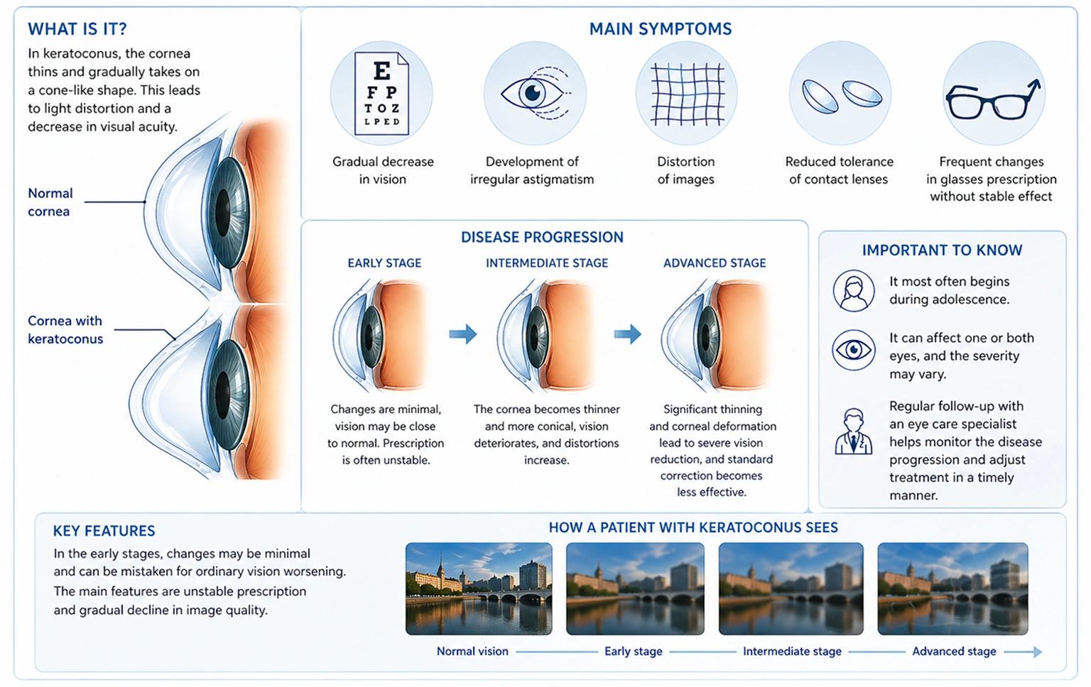
Keratoconus most often begins in adolescence, during a period of rapid growth, and can remain undetected for a long time. In the early stages, corneal changes are minimal, so patients don’t always seek medical attention immediately.
The disease can affect one or both eyes, and the severity of the changes often varies. In most cases, the process develops gradually, leading to a gradual deterioration in vision.
Main symptoms:
- gradual vision loss
- development of irregular astigmatism
- image distortion
- decreased tolerance of contact lenses
- frequent changes in spectacle correction without a stable effect
In the initial stages, changes may be minimal and perceived as normal vision loss. However, a characteristic feature is instability of correction and a gradual decline in image quality.
As the condition progresses, the cornea becomes increasingly thinner and deformed, leading to significant vision loss and a decrease in the effectiveness of standard correction.
Diagnosis and Signs
The most sensitive method for early diagnosis is corneal topography, which allows for the detection of changes in corneal shape even before the onset of significant symptoms. This test provides a detailed map of the corneal surface and allows for the detection of even minimal deviations from the norm.
In the early stages, instrumental diagnostics play a key role, as clinical manifestations may be absent or mild.
As the disease progresses, characteristic clinical signs may appear:
- Fleischner’s ring — iron deposition in the epithelium around the cone
- Munson’s sign — protrusion of the lower eyelid when looking downward
- Cicatricial changes in the cornea in severe cases
If Descemet’s membrane ruptures, corneal edema may develop, accompanied by a sharp deterioration in vision.
Modern diagnostics also include assessment of the thickness and biomechanical properties of the cornea, which allows for the earliest possible detection and prompt initiation of treatment.
Forms and Degrees of the Disease
There are three morphological types of keratoconus, distinguished by the size and location of the corneal deformity:
- Mammillary – up to 5 mm in diameter, with a pronounced steepness and a centrally or paracentrally located apex
- Oval – larger, with a downward and laterally deviated apex
- Spherical – more than 6 mm, involving a significant portion of the cornea
This classification is important for assessing disease progression and choosing the optimal treatment strategy.
By severity, the following are distinguished:
- Initial – less than 48 diopters
- Moderate – 48–54 diopters
- Severe – more than 54 diopters
The severity of the disease directly impacts vision quality, the effectiveness of correction, and the choice of treatment method. In the early stages, keratoconus can be virtually asymptomatic and only detected through instrumental diagnostics. The transition between stages occurs gradually, requiring regular monitoring and follow-up examinations to assess the progress of changes.
Disease Progression
Keratoconus typically progresses slowly, but in some cases, spontaneous stabilization is possible. The rate of change can vary and depends on the patient’s age, individual corneal characteristics, and the presence of associated factors.
The main problem is that standard spectacle correction becomes insufficient because the cornea’s shape changes unevenly. This leads to vision instability and reduced image quality, even with properly fitted glasses.
This particular feature requires an individualized approach to vision correction and ongoing monitoring by a specialist to assess the progress of the disease. Regular examinations allow for timely detection of changes and the adjustment of treatment strategies.
Modern Treatment Methods
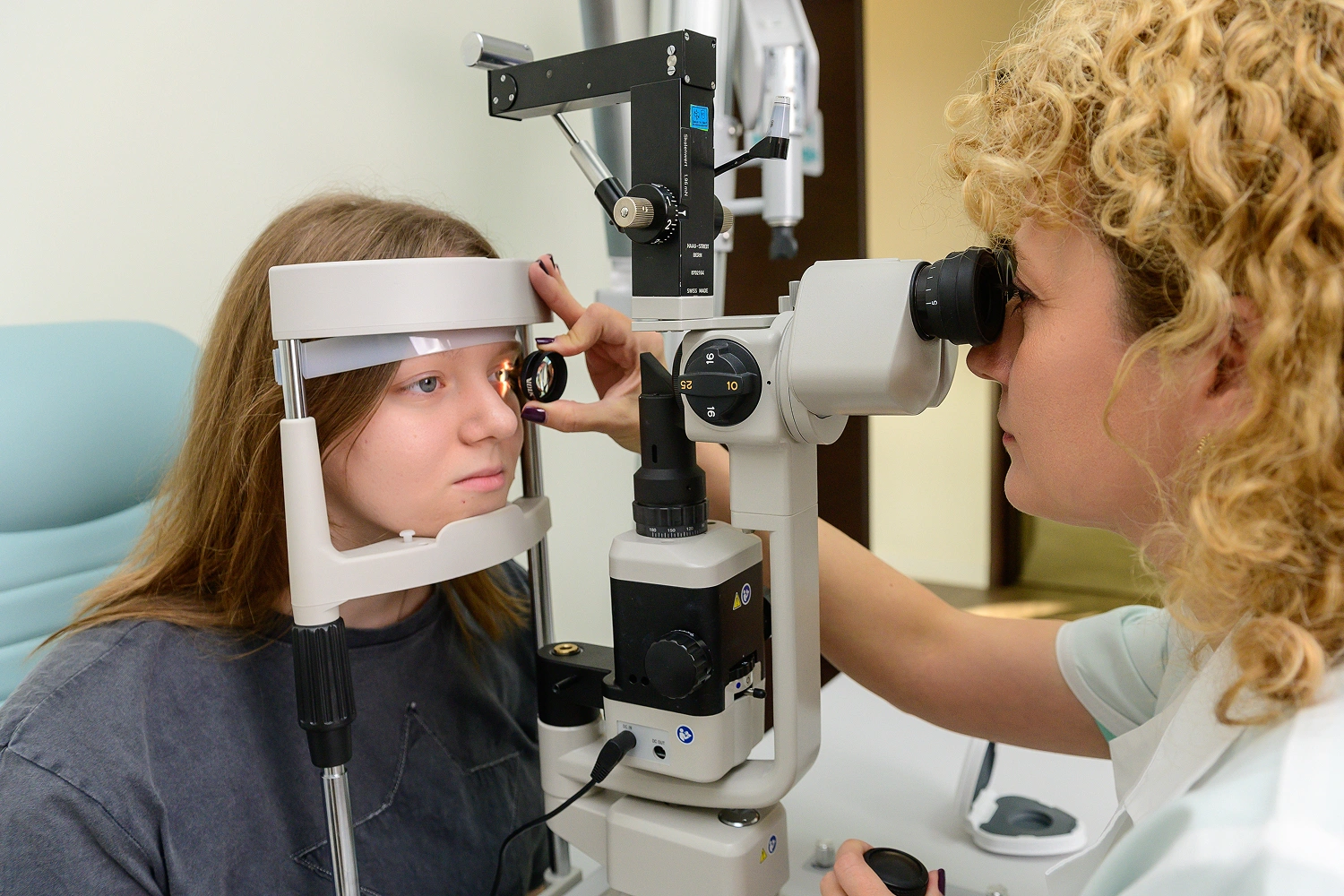
Keratoconus treatment strategies are determined by the stage of the disease, the severity of changes, and the rate of their progression. Modern practice utilizes a step-by-step approach that not only improves vision but also stabilizes the cornea.
The main goal is to preserve its structure, slow the progression of the disease, and select the most effective correction method at each stage.
1. Conservative Correction
In the early stages, the following are used:
- rigid gas-permeable lenses
- scleral lenses
These lenses create a stable optical surface, compensating for the irregular shape of the cornea and significantly improving vision.
Unlike soft lenses, they provide more precise correction for irregular astigmatism and allow for predictable results.
Selection is individualized based on a detailed diagnosis of corneal parameters, including its shape, thickness, and surface characteristics.
2. Corneal Crosslinking
Crosslinking is a key method aimed at strengthening the corneal collagen framework and increasing its mechanical stability.
The procedure includes:
- epithelial removal
- application of a riboflavin solution
- exposure to ultraviolet radiation
This results in the formation of additional bonds between collagen fibers, which helps slow or stop the progression of the disease.
The most pronounced effect is achieved in the early and middle stages, when the corneal structure is still preserved.
3. Intrastromal segments
Implantation of corneal segments is used for more pronounced changes in the shape of the cornea.
The segments are inserted into the cornea and change its geometry, reducing the degree of deformation and improving optical characteristics.
This method is used according to indications and is often combined with other treatments, allowing for a more stable and controllable result.
4. Surgical treatment
As the disease progresses and vision significantly decreases, the following may be required:
- penetating keratoplasty (corneal transplant)
This method is used in cases where conservative and minimally invasive approaches fail to provide the desired effect. The surgery aims to restore corneal structure and is tailored to the individual patient’s needs.
Modern ophthalmology emphasizes early diagnosis and timely intervention, which helps slow the progression of keratoconus and preserve vision without radical surgery.
Combined approaches—crosslinking, contact lens correction, and minimally invasive techniques—allow for individualized treatment for each patient.
This approach not only helps manage the progression of the disease but also significantly improves visual function.
Why is it important not to delay diagnosis and treatment
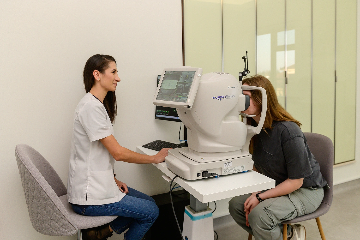
Keratoconus is a progressive corneal disease, so early detection is key to preserving vision. In the early stages, changes may be minimal, but it is during this period that treatment produces the most significant and stable results.
With the right approach, it can not only halt the progression of the disease but also significantly improve vision without the need for radical interventions.
Regular follow-up with a specialist allows us to monitor the dynamics of changes, promptly adjust treatment, and select the most effective methods at each stage.
Consultation and Treatment at Visus Clinic
Learn more about modern methods for diagnosing and treating keratoconus at Visus Clinic and schedule a consultation.
We perform detailed diagnostics, including keratotopography and corneal assessment, allowing us to accurately determine the stage of the disease and select the optimal treatment strategy.
Our goal is not only to improve vision but also to halt the progression of the disease, preserving vision for years to come.
Answers to Patient Questions
- Is the disease treatable?
Yes, modern methods can slow or stop the progression and improve vision. - Is it possible to fully restore vision?
It depends on the stage. In the early stages, significant improvement is observed; in later stages, the main goal is stabilization and vision correction. - Is crosslinking painful?
The procedure is performed under local anesthesia and is generally well tolerated by patients. - Is surgery necessary?
No. In the early stages, contact lenses and regular monitoring are often sufficient. Surgical treatment is used when the condition progresses and significant changes occur. - How quickly does the disease progress?
The rate varies from person to person. In most cases, the process is slow, but regular monitoring is required. - Can I wear contact lenses?
Yes, rigid and scleral lenses are one of the main methods of vision correction for this condition. - Is surgery possible?
In the early stages, this is possible with timely use of modern treatment methods and specialist monitoring. - When should I see a doctor?
At the first signs of deteriorating vision or instability of correction, it is recommended to undergo diagnostics as early as possible.